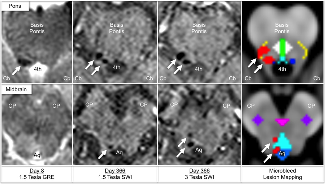
Fig. 2.

Mapping traumatic microbleeds in the brainstem. Traumatic microbleeds are punctate hypointense lesions indicated by white arrows in the rostral pons (top row) and caudal midbrain (bottom row). In the right panel, traumatic microbleeds identified by 3 Tesla susceptibility-weighted imaging (SWI) are coregistered to Montreal Neurological Institute (MNI152) space and superimposed upon a template of brainstem arousal nuclei. Traumatic microbleeds are colored red, and brainstem arousal nuclei are color-coded as follows: dark blue, locus coeruleus; turquoise, dorsal raphe´; white, pontis oralis (pontine reticular formation); green, median raphe´; yellow, parabrachial nuclear complex; light blue, periaqueductal grey matter; pink, ventral tegmental area; purple, pedunculopontine nucleus. The traumatic microbleeds overlap partially with the right-sided locus coeruleus, parabrachial nuclear complex, dorsal raphe´, and periaqueductal grey matter. Neuroanatomic landmarks: Cb, cerebellum; 4th, fourth ventricle; CP, cerebral peduncle; Aq, cerebral acqueduct. Of note, although the GRE and SWI datasets were acquired on different days, the paramagnetic properties of blood are expected to produce the same hypointense signal at each time point.
