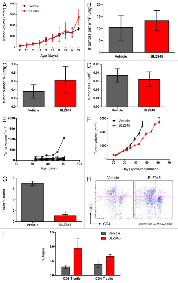
Figure 3. Pharmacological blockade of CSF1R signaling increases infiltration of T cells and decreases tumor growth but does not affect pulmonary metastasis in PyMT mice. (A–E) 63- to 70-d old MMTV-PyMT transgenic mice were randomized by total tumor burden and dosed with 200 mg/kg BLZ945 or vehicle (n = 9 per group) at the indicated time points. Individual tumor volumes were calculated by caliper measurements with total tumor burden being the sum of these measurements. (A) Cumulative tumor burden of vehicle- and BLZ945-dosed mice. (B) Lungs from MMTV-PyMT mice were formalin-fixed and serially sectioned to histologically evaluate the number of individual metastases per mm2 of lung. (C) The total metastatic tumor area as a percentage of lung tissue. (D) The average area (mm2) of lung metastatic spread (E) Representative graph showing individual tumor volumes taken from a vehicle-treated mouse. (F–I) Spontaneous tumors from naïve MMTV-PyMT mice were pooled and digested to form a single-cell suspension. Cells were injected into mammary fat pads of syngeneic mice. PyMT allograft-recipient mice with average tumor volumes ~280 mm3 were randomized into 2 groups and dosed with 200 mg/kg BLZ945 or vehicle control 21 d post-implantation. (F) Caliper measurements of tumor volumes (n = 6 per group) were taken every 3–4 d. In a separate study, tumors (n = 4 per group) were analyzed by flow cytometry to determine infiltration of (G) CD45+CD11b+Ly-6G/C(Gr-1)−/loF4/80+ TAMs and (H and I) CD45+CD3+CD4+ and CD45+CD3+CD8+ T cells in tumor allografts . Graphs display mean values ± SEM. Statistical analyses were performed by 2-tailed unpaired Student t test; *P < 0.05 vs. vehicle; data shown are representative of at least 2 experiments.

An official website of the United States government
Here's how you know
Official websites use .gov
A
.gov website belongs to an official
government organization in the United States.
Secure .gov websites use HTTPS
A lock (
) or https:// means you've safely
connected to the .gov website. Share sensitive
information only on official, secure websites.
