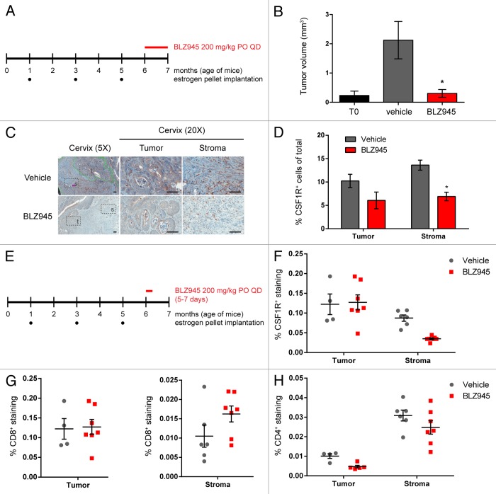
Figure 4. Treatment with BLZ945 reduces macrophages, enhances T cell infiltration, and prevents tumor growth in the K14-HPV16 transgenic mouse model of cervical carcinoma. (A–D) Estrogen pellets were administered to 1-mo old K14-HPV16 transgenic mice every 2 mo. Age-matched control mice that did not receive estrogen pellets are referred to as “T0.” At 6 mo of age, mice were dosed with 200 mg/kg BLZ945 or vehicle control for 1 mo, after which time whole cervixes were formalin-fixed for histological analyses. (B) Serial sections of cervical tissue were hematoxylin and eosin (H&E) stained and tumor volumes determined by multiplying the tumor area by the depth of serial sections. Cervical tumors from 6-mo old estrogen-naïve mice (T0) served as a baseline control. (C and D) Cervical tissues were stained with an anti-CSF1R antibody to label macrophages by immunohistochemistry. Magnified views of tumor (t) and stroma (s) regions within the cervix are boxed. Scale bar, 100 µm. (D) Quantification of CSF1R staining in cervical tumor and stroma regions. (E–H) Pharmacodynamic study to monitor changes in tumor-infiltrating and stromal immune cells after 5–7 d of treatment with BLZ945. Whole cervixes were frozen, serially sectioned, and stained with H&E to identify the transformation zone. Tumor and stroma regions within the cervix were scored separately for (F) CSF1R+ macrophages, (G) CD8+ T cells, and (H) CD4+ T cells. Bar graphs represent mean values with SEM. Statistical analyses were performed by Mann–Whitney nonparametric test; *P < 0.05 vs. vehicle.

An official website of the United States government
Here's how you know
Official websites use .gov
A
.gov website belongs to an official
government organization in the United States.
Secure .gov websites use HTTPS
A lock (
) or https:// means you've safely
connected to the .gov website. Share sensitive
information only on official, secure websites.
