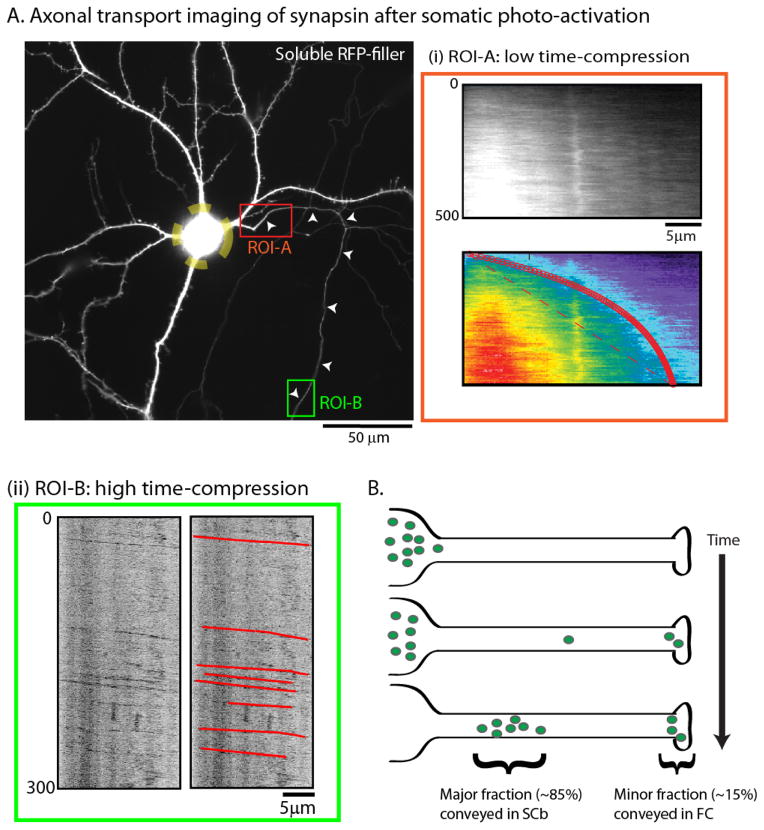
Figure 4. A fraction of SCb proteins are conveyed in the fast component.
(A) To more closely simulate the radiolabeling paradigm, cultured neurons were transfected with GFP:synapsin and soluble mRFP (shown); the neuronal soma was photoactivated (yellow dashed ROI); and the egress of photoactivated molecules into the emergent axon was evaluated over time. While the bulk of synapsin molecules moved slowly into the proximal axon with kinetics expected for slow axonal transport (ROI-A – red box), rapidly-moving particles of synapsin were also seen when the distal axon was imaged after somatic photoactivation (ROI-B – green box).
(B) The “dynamic recruitment” model for SCb transport. After synthesis in the soma, cytosolic molecules intermittently and probabilistically associate with “carriers” moving in fast axonal transport. As such, some molecules remain associated with these carriers for long periods, giving rise to a small population (~ 10–15%) that is rapidly transported to axons and synapses. However the majority of cytosolic molecules are slowly conveyed with kinetics resembling slow axonal transport. An implication of this model is that common transport “carriers” are responsible for conveying both fast component and SCb proteins. Figure adapted from Scott et al., 2011, with permission.

