Figure 1.
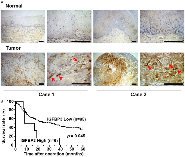
High IGFBP3 expression is associated with poor prognosis in ESCC patients. A: Immunohistochemistry reveals focal upregulation of IGFBP3 in primary ESCC tissues on tissue microarrays. Two representative cases scored as strongly positive IGFBP3 expression in tumors are shown. Note an intense cytoplasmic staining of IGFBP3 (arrows). IGFBP3 was expressed in spindle-shaped tumor cells reminiscent of EMT in case 1. Scale bars, 50 μm. B: A high level of IGFBP3 expression predicts a poor 5 year survival rate. Overall survival curves were plotted according to the Kaplan-Meier method, and p value was calculated using log rank test.

