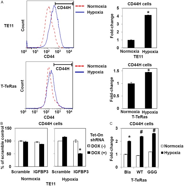
Figure 4.
IGFBP3 mediates hypoxic induction of CD44H cells in culture. TE11, T-Te-Ras and indicated derivatives were exposed to hypoxia (0.5% O2) or normoxia for 48 h and analyzed by FACS for CD44H cells. In (A), a segmented line indicates the CD44H cell fraction. In (B), TE11 cells with Tet-On shRNA directed against IGFBP3 or a non-silencing sequence (scramble) were treated in the presence or absence of 0.5 μg/ml DOX. In (C), Bla, empty vector control; WT, wild-type IGFBP3; GGG, GGG-mutant IGFBP3. *, P<0.05 vs. Normoxia (n=3) in (A); P<0.05 vs. Scramble and DOX (+) (n=3) in (B); and P<0.05 vs. Bla and Normoxia (n=3) in (C). #, P<0.05 vs. Bla and Hypoxia (n=3) in (C).

