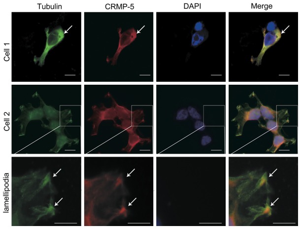
Figure 2.
Colocalization of CRMP-5 and tubulin in HEK293 cells. Anti-tubulin and anti-CRMP-5 were used to detect endogenous tubulin and CRMP-5 proteins in HEK293 cells; DAPI was used to stain the nuclei. The merged images show the colocalization (yellow spots) of tubulin (green) with CRMP-5 (red). Enlarged images of cells show details of the lamellipodia. Scale bar, 20 μm.

