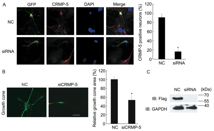
Figure 4.

CRMP-5 is necessary for growth cone development in cultured hippocampal neurons. A: Hippocampal neurons cultured 48 h were co-transfected with scrambled siRNA (NC) or CRMP-5-siRNA together with a GFP-encoding plasmid; after 24 h, the neurons were fixed and subjected to the immunocytochemistry protocol. Endogenous CRMP-5 was detected by an anti-CRMP-5 antibody. Typical images of the CRMP-5 immunostaining are shown (left panel). A non-targeting siRNA was used as the negative control (NC). The percentages of CRMP-5-positive neurons with each treatment were quantified (right panel) as the mean ± SEM for three independent experiments. *Denotes P<0.05; Scale bar, 10 μm. B: Typical growth cone morphology in neurons with each treatment (left panel). The growth cone area of transfected cells was determined and plotted. Comparisons were made using one-way ANOVA. *Denotes P<0.05 (n=30-40 cells from 3 independent experiments). Error bars indicate SEM. Scale bar, 10 μm. C: HEK293 cells were co-transfected with scrambled siRNA (NC) or siCRMP5 together with the FLAG-CRMP-5 expression plasmid. Lysates were probed with anti-CRMP5 antibody. The GAPDH antibody was used as a loading control.
