Figure 1.
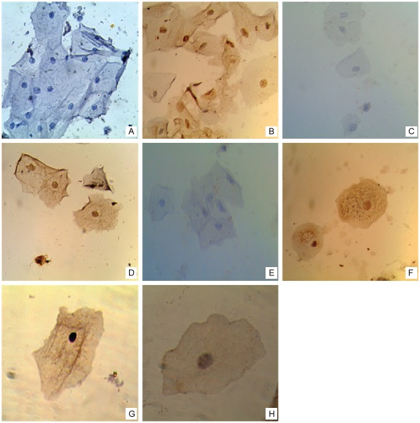
Expression of E6, p53 and 21, and physical state of HPV16 genome in cervical smears. Representative images of immunocytochemical staining for E6 (A, B), p53 (C, D) and p21 (E, F) proteins (40X). (A) Negative in Non-IL and (B) positive immunostaining for E6 in LSIL. (C) Negative and (D) positive immunostaining for p53 in Non-IL. (E) Negative and (F) positive immunostaining for p21 in LSIL. (G, H) Representative images of in situ hybridization for HPV16 genome (100X). (G) Diffuse signal pattern in Non-SIL, and (H) punctate signal pattern in LSIL.

