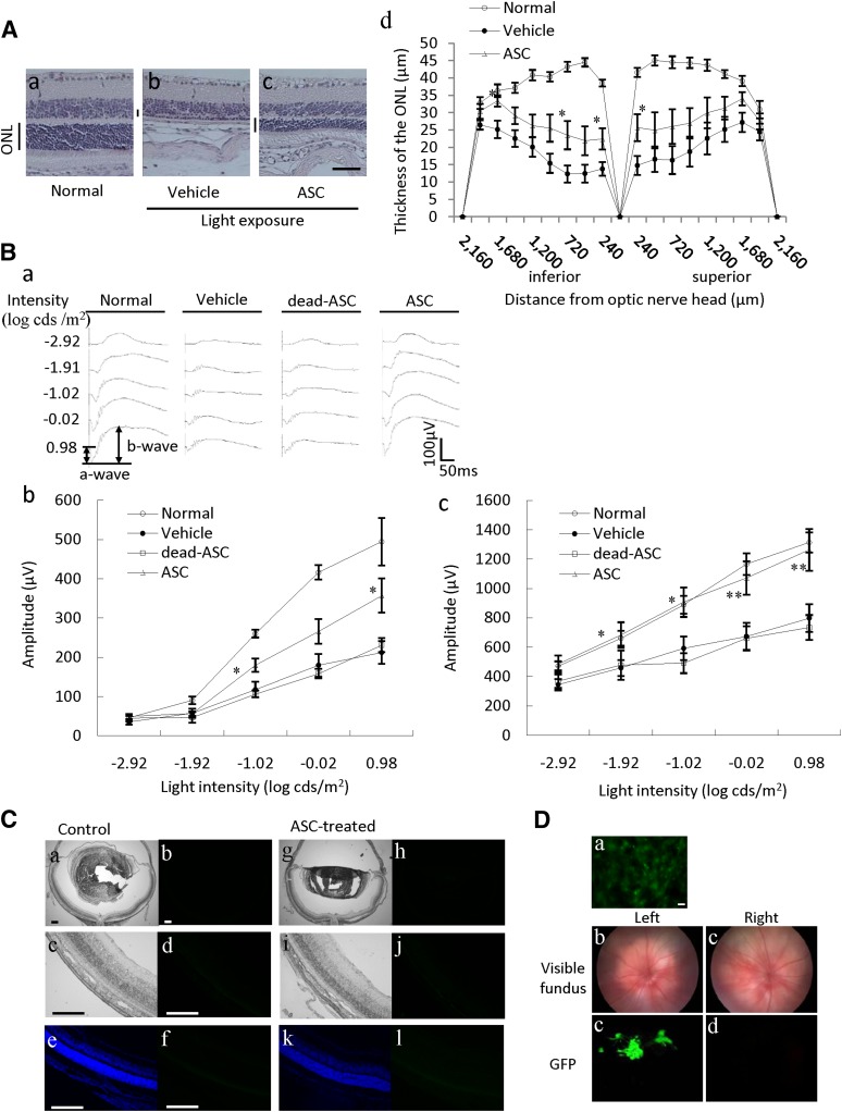
Figure 1.
ASCs reduced retinal damage induced by exposure to light in mice without engraftment. (A): Light-induced retinal degeneration was reduced by ASCs. Representative photographs of hematoxylin and eosin staining are as follows: nontreated group (Aa), light exposure (8,000 lx) plus vehicle-treated group (Ab), and light exposure plus ASC-treated (103 cells per eye, intravitreal administration) group (Ac) at 28 days after light exposure in mice. (Ad): Thickness of the ONL was measured at 28 days after light exposure. The ONL was measured at 240-μm intervals from the optic disc. Scale bar = 50 µm. (B): Light-induced retinal dysfunction was reduced by ASCs. (Ba): Typical traces of dark-adapted electroretinogram responses measured at 28 days after exposure to light. Stimulus flashes were used from −2.92 to 0.98 log-candela seconds/m2. Amplitudes of a- and b-waves of the light exposure (8,000 lx) plus vehicle-treated group (Bb) versus the light exposure plus ASC-treated group (103 cells per eye) group (Bc) are shown. (C): ASCs were not engrafted into the retina. (Ca–Cf): Nontreated. (Cg–Cl): ASCs were injected into the vitreous chamber. (Ca, Cc, Cg, Ci): Images of retinal cross-section. (Cb, Cd, Ch, Cj): Bright-field images. (Ce, Cf, Ck, Cl): Fluorescence images to detect GFP transgenic mouse-derived ASCs. Images of retinal cross-sections showing Hoechst staining (Ce, Ck) and immunostaining with GFP antibody (Cf, Cl). Scale bars = 200 µm. (D): Fluorescence of CAG-EGFP mouse-derived ASCs. (Da): The cultured ASCs were investigated using a fluorescence microscope. Scale bars = 50 µm. The ASCs were injected into the left vitreous, and the ocular fundus (Db, Dc) and the fluorescence of ASCs in the vitreous body (Dd, De) were investigated using a fluorescence retinal microscope. Data are shown as mean ± SEM, n = 7 or n = 8. *, p < .05 versus the light exposure plus vehicle-treated group (Student’s t test). Abbreviations: ASC, adipose-derived stem cell; GFP, green fluorescent protein; ONL, outer nuclear layer.

