Figure 4.
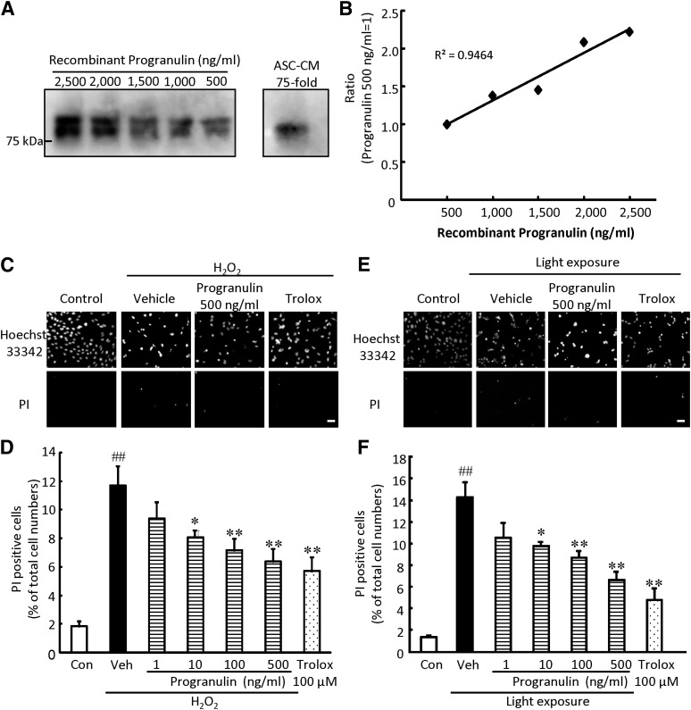
Progranulin suppressed H2O2- or light-induced cultured photoreceptor cell death. (A, B): Quantitative determination of progranulin in ASC-CM by Western blot. (A): Representative immunoblots showing progranulin protein levels in ASC-CM and recombinant mouse progranulin. The recombinant progranulin was expressed in a mouse myeloma cell line, and post-translational modifications such as glycosylation may contribute to various molecular weights. (B): Standard curve was generated from the density of mouse recombinant progranulin. (C, E): Representative fluorescence microscopy images showing nuclear staining for Hoechst 33342 and PI after 27 hours of H2O2 (0.3 mM) treatment (C) or 24 hours of light exposure (E). (D, F): The number of cells exhibiting PI fluorescence was counted, and positive cells were expressed as the percentage of PI- to Hoechst 33342-positive cells. The number of PI-positive cells increased after H2O2 treatment or light exposure. Progranulin significantly reduced cell death in a concentration-dependent manner. Scale bars = 50 µm. Data are shown as mean ± SEM (n = 6 or n = 9). *, p < .05 versus vehicle; **, p < .01; ##, p < .01 versus control. Abbreviations: ASC-CM, adipose-derived stem cell-conditioned medium; Con, control; H2O2, hydrogen peroxide; PI, propidium iodide; Veh, vehicle.

