
Figure 3.
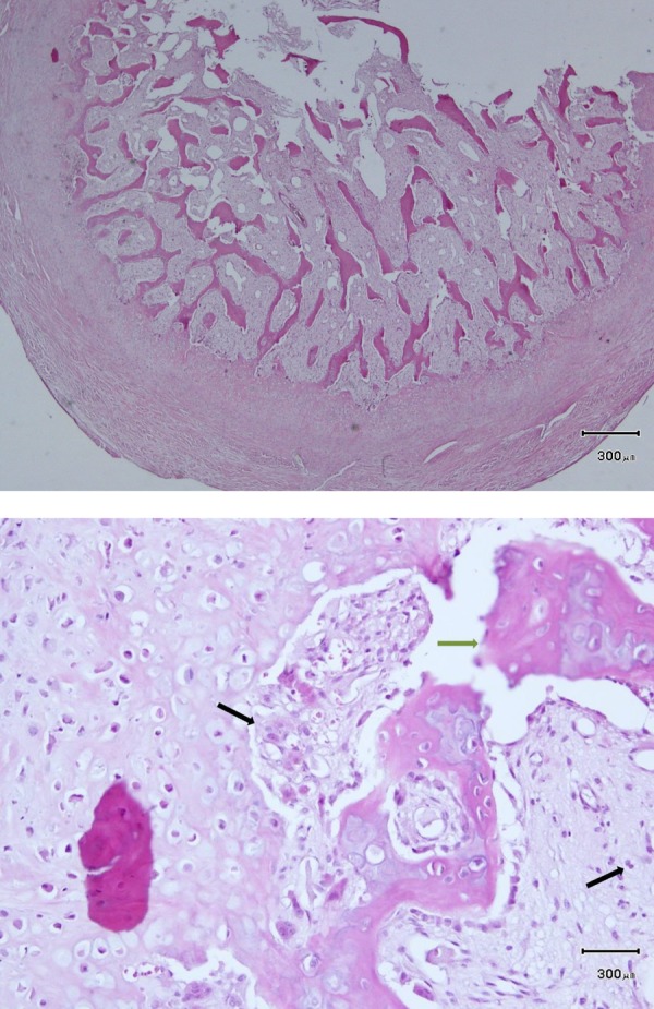
(A) Photomicrographs showing a hypercellular cartilaginous cap with underlying bone trabeculae and intertrabecular fibroblastic proliferation (H&E ×100). (B) A higher magnification photomicrograph showing hypercellular cartilage and areas of calcification in the cartilaginous cap (H&E ×400).