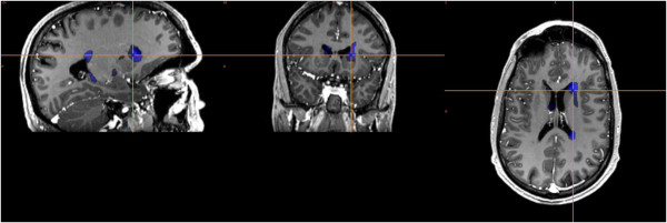
Figure 2.
Computer-aided subtraction SPECT imaging in a patient with severe dystonia. The patient was 35 years old with long history of cervical dystonia and choreoathetotic movements. We obtained Technetium 99 m Neurolite SPECT scan of the brain in this patient with awake state and under anesthesia, respectively. Subtraction SPECT imaging had shown a hypermetabolic left caudate (blue), which is helpful for DBS target selection.

