Figure 1.
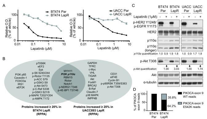
Lapatinib-resistant cells feature enhanced PI3K p110α activation. (a) BT474 and UACC893 parental (Par) and LapR cells were treated for 4 days (BT474) or 3 days (UACC893) as shown followed by MTT assay. (O.D., optical density, is an indicator of cell number.) Results are representative of two independent experiments. Error bars, s.e.m. (b) Reverse phase protein array (RPPA) analysis was performed on BT474 and UACC893 parental and LapR cells. Proteins increased ≥20% in LapR versus parental cells are shown. (c) Cells were treated 4.5 hours (BT474) or 4 hours (UACC893) with DMSO or 1μM lapatinib, followed by western blot. (d) Whole-exome sequencing was performed on genomic DNA and percent of WT and mutant reads at position corresponding to codon 542 in p110α (PIK3CA gene) were compared in BT474 parental versus LapR cells.

