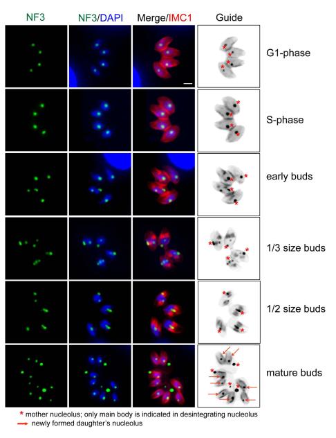
Figure 4. Major morphogenic changes of the nucleolus during tachyzoite replication.
Immunofluorescence of factor TgNF3 reveals the tachyzoite nucleolus undergoes dramatic morphogenic changes during parasite division. Nucleolus structure and organization was examined in replicating 11-104A4 parasites (grown at 34°C) first synchronized by limiting invasion (see Experimental Procedures) and then inoculated into cover slips for IFA analysis. At various times, samples were harvested and co-stained with antibodies for TgNF3 and IMC1 as well as stained with DAPI. Representative nucleolar morphology at six cell cycle phases and transitions is shown as identified by established cell cycle criteria based on parasite size and shape (red=IMC1 staining), nuclear morphology (DAPI, single versus double nuclei), and the presence or absence of internal daughters (also red=IMC1). Note immediately following the initiation of budding (early buds), the nucleolus began to fragment with some nucleolar material occasionally lost in the mother cell residual mass. The stars in the marker guide panel (B&W inverse of merged IMC1 and TgNF3 stains), indicates nucleolar material, while the arrows in the bottom guide image indicate the nucleoli of new daughter parasites. Scale bar = 2μm.

