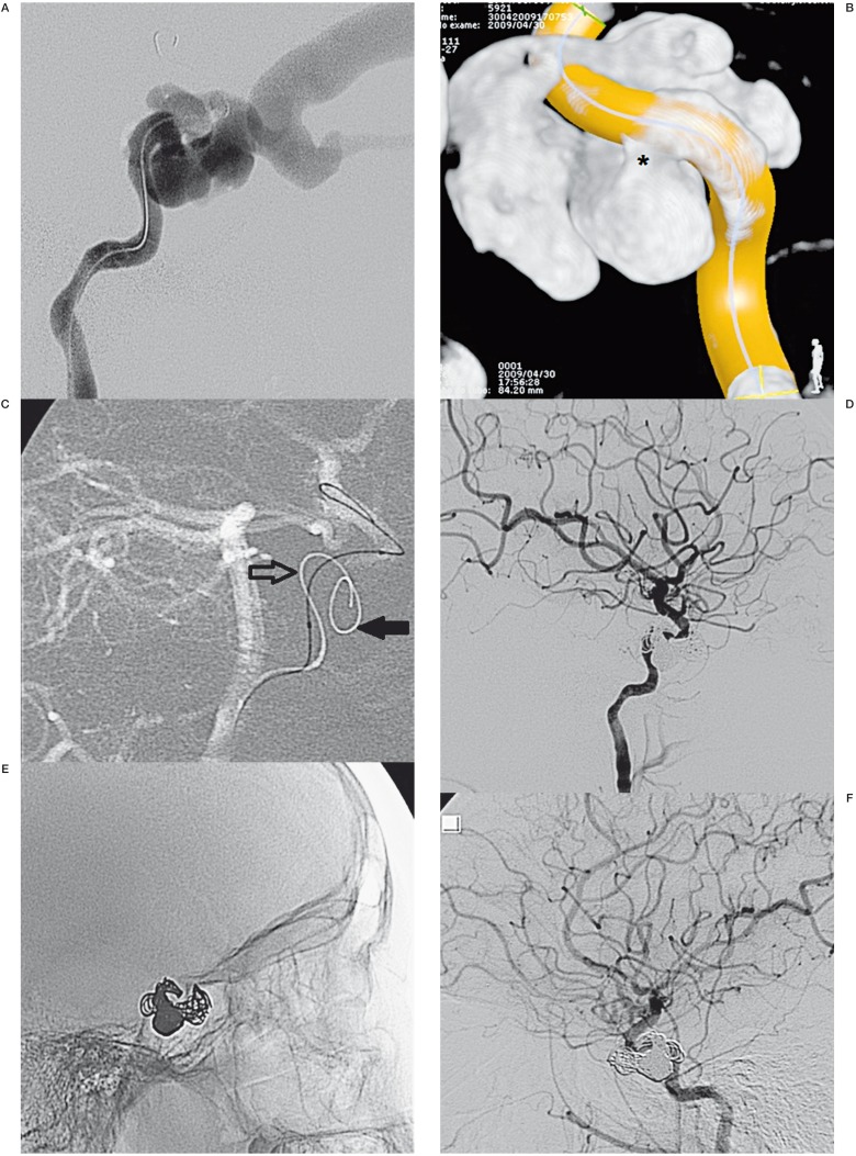
Figure 2.
A) Lateral view of a left ICA angiogram showing a high-flow CCF draining into the left superior and inferior ophthalmic vein. B) Rotational acquisition demonstrates the exact location of the fistula (asterisk). C) Lateral view of the left ICA angiogram reveals two microcatheters with the first being inserted into the cavernous sinus (filled arrow) and the balloon along the internal carotid (open arrow). D,E) Immediate angiograms confirmed a complete obliteration of the fistula with 12 coils. F) Three months after the angiogram, a complete resolution of the CCF was observed. ICA, internal carotid artery; CCF, carotid cavernous fistula.

