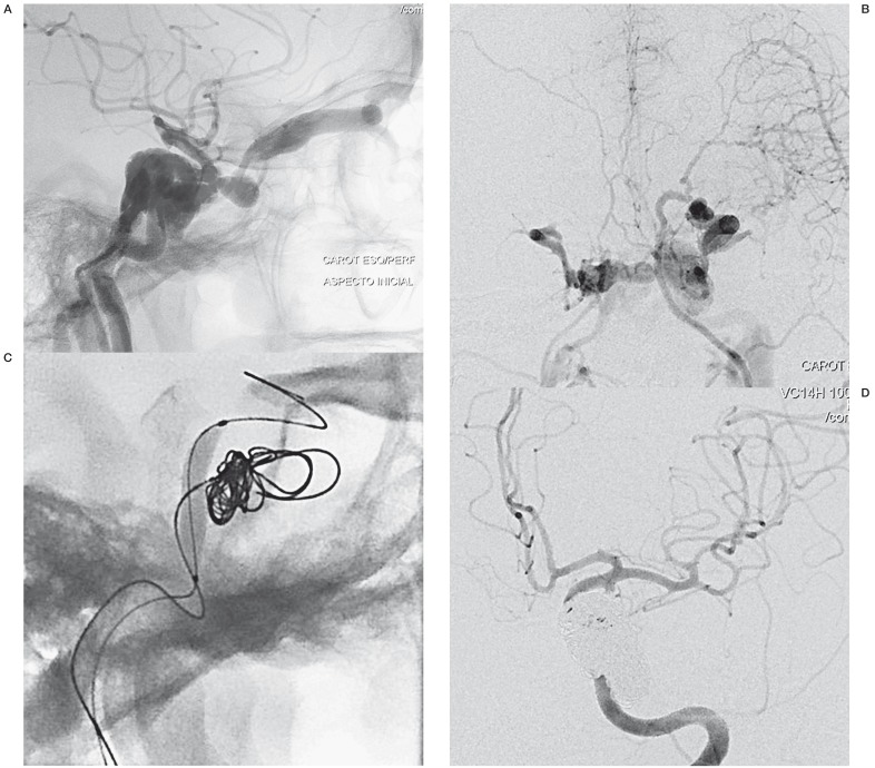
Figure 4.
A) Lateral view of a left ICA angiogram showing a high-flow CCF draining into the left superior ophthalmic vein and inferior petrosal sinus. B) Anterior-posterior view in the venous phase, showing CCF draining into the contralateral CS. C) Angiographic view of the compliant balloon and a cast of coils. D) Three months later, a control angiogram revealed a complete resolution of the CCF. ICA, internal carotid artery; CCF, carotid cavernous fistula; CS, cavernous sinus.

