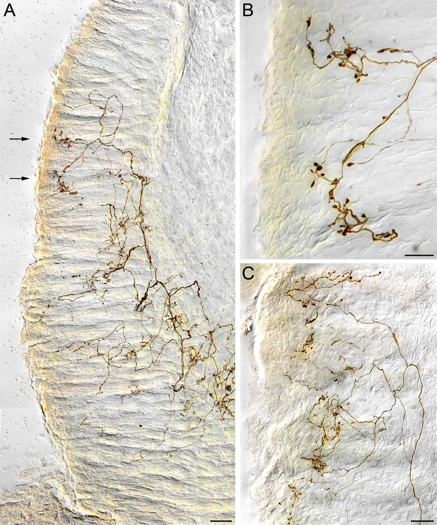
Figure 5.
Vagal antral gland afferents. A: A low-power view of part of the terminal arbor of an afferent innervating the gastric mucosa. One of two second-order collaterals of a vagal afferent fiber (entering from lower right corner of plate) distributes to form an arbor of higher order processes that travel along the glandular epithelial walls of the antral mucosa immediately adjacent to the antral lumen (far left of image). The collaterals tend to form varicose terminals and in some cases lamellar processes along the basal surfaces of the epithelial walls of the antral glands. On reaching the epithelium that constitutes the luminal surface, the collaterals often form aggregates of terminal varicosities and swellings. B: A higher-power image of two luminal aggregates of varicosities taken from the case illustrated in panel A (the area of the aggregates is designated with arrows in panel A). C: A lower-power image illustrating part of the arbor of terminals of a collateral of an antral afferent. In this case, the afferent collateral can be seen bifurcating in the lamina propria before coursing into the glandular mucosa to arborize. Scale bars = 50 µm in A; 20 µm in B; 25 µm in C.

