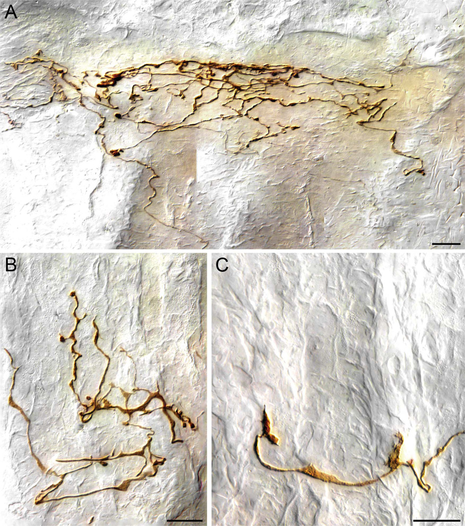
Figure 6.
Vagal antral afferent terminals in the gastric mucosa. A: An antral afferent neurite travels (from bottom of panel) to arborize into a plate of terminals immediately below the luminal epithelium of the antrum (in this case, the lumen is just immediately above the top of the image, and the gastric glands are running vertically to secrete into the lumen). B: An aggregate of terminals of an antral afferent located within the glandular wall. In this example, and in all panels of this figure, the glands are oriented in a vertical direction, and the afferent terminals are distributed among the necks of the glands, not at the gastric lumen. As illustrated by this terminal, many of the deeper terminal endings of the antral afferents–particularly those elements of the neurite that coursed laterally or at right angles to the length of the gastric glands–were commonly lamelliform or flattened in appearance. C: An example of the lamellar or growth-cone shaped terminals often formed by the antral afferents. Scale bars = 20 µm in A; 15 µm in B,C.

