Fig. 1.
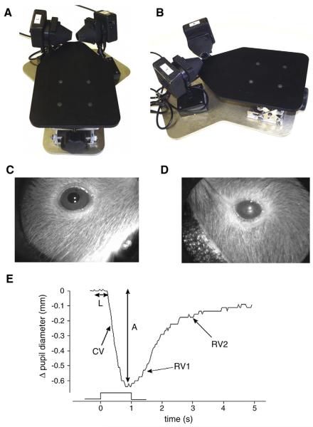
A) Mouse pupillometer top-frontal view and B) top-lateral view. During the examination, the mouse sits in the center of the platform (not shown) with two adjustable pupil cameras focused on the eyes. Panel C) shows the amplified view of a healthy murine pupil, whereas panel D) depicts a C57BL/6 mouse with cataract. Similar observations were made in other mouse strains. Direct and consensual pupillary responses were mostly reproducible in mice with cataracts. However, light reflections from the clouded crystalline lenses commonly made it difficult to distinguish the outer border of the pupil from the iris. E) Example of mouse pupil light reflex responses indicating measurement of key autonomic parameters. The trace represents a normal pupil response to a 1s light flash. Onset latency (L) and response amplitude (A) are measured as shown. Constriction velocity (CV) represents the maximal downward slope of the response curve. The biphasic re-dilation response is shown with the initial re-dilation velocity (RV1) and slower second re-dilation velocity (RV2).

