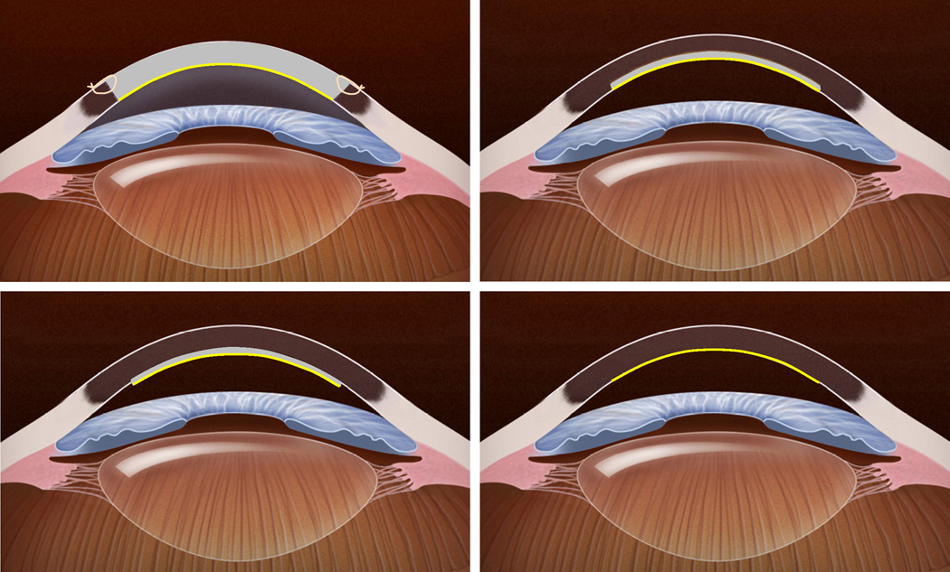
Figure 1. Schematic representation of different keratoplasty techniques for endothelial disease.
Top Left, In penetrating keratoplasty, the full-thickness host cornea is replaced by a full-thickness donor cornea to provide donor endothelial cells (yellow). Sutures are required to secure the donor tissue to the host. Top Right, In deep lamellar endothelial keratoplasty, the posterior host corneal stroma, Descemet membrane and endothelium are excised through a large or small limbal incision and replaced with donor tissue consisting of similar layers. This provides donor endothelial cells (yellow) without corneal incisions or sutures, but introduces an irregular, manually dissected lamellar interface between the donor tissue and host corneas. Bottom Left, In Descemet stripping endothelial keratoplasty, host Descemet membrane and endothelium are stripped from the host posterior corneal stroma, and replaced with donor tissue consisting of posterior stroma, Descemet membrane and endothelium. This provides donor endothelial cells (yellow) without an anterior corneal incision or corneal sutures. Because the donor tissue is usually prepared with a mechanical microkeratome, and the posterior stromal surface is not disrupted, the lamellar interface between donor tissue and host is regular, but the procedure adds a variable thickness of stromal tissue. Bottom Right, In Descemet membrane endothelial keratoplasty, host Descemet membrane and endothelium are stripped from the host posterior corneal stroma, and selectively replaced with donor Descemet membrane and endothelium (yellow). This anatomically mimics the native cornea without requiring an anterior corneal incision, but manipulation of the thin, delicate donor membrane can be technically challenging.

