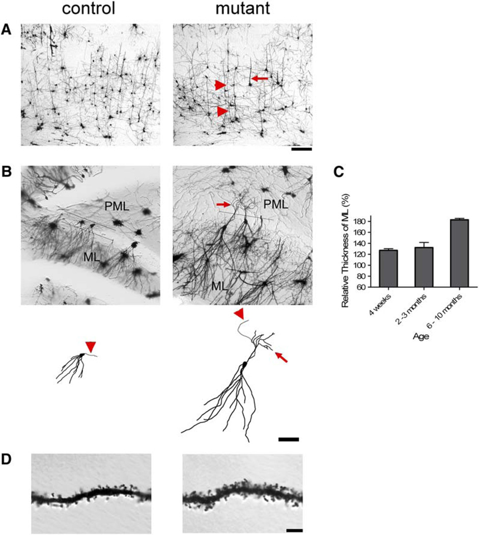
Figure 6. Golgi Stain Revealed Dendritic Hypertrophy, Ectopy, and Increased Spine Density in Pten-Deleted Brain.
(A) Thickened or elongate neuronal processes (arrow and arrow heads, respectively) were present in mutant cerebral cortex compare to control at 3 months of age. Scale bar, 100 µm.
(B) At 8 months of age, increased length of the dendritic arbors in the molecular layer (ML) and ectopic neuronal processes (arrow) in the polymorphic layer (PML) were observed in mutant compare to control (upper panels). Reconstructions of single neurons made from image stacks emphasize the dendritic hypertrophy of the mutant neurons compare to control (lower panels). An axon can be seen emanating from both mutant and control neurons (arrowheads). Scale bar, 100 µm.
(C) Mutant ML was significantly thicker than that of control at all adult ages tested (p < 0.05).
(D) Higher-magnification images of dendrites in the ML revealed increased thickness and spine density in mutant (1.434 ± 0.064 spines/µm) versus control (1.077 ± 0.033 spines/µm) brains (p < 0.000005, n = 23 and 26 dendritic branches from three brains, respectively). Scale bar, 10 µm.

