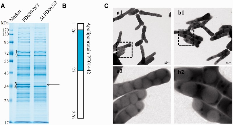
Figure 5.
Deletion of LPD06283 results in supersized LDs. (A) Gel electrophoresis of LD proteins from R. opacus PD630-WT and the LPD06283 deletion mutant stained by colloidal blue. Band 3, the main band, disappeared in the LPD06283 deletion mutant. (B) Location of the predicted domain in LPD06283, Apolipoprotein (PF01442). (C) a1-a2, EM images of R. opacus PD630-WT cultured in MSM for 24 h after growing in NB for 48 h using positive staining methods; b1-b2, EM images of the LPD06283 deletion mutant under the same conditions as R. opacus PD630-WT. The lower panels give amplified pictures. Bar = 2 μm.

