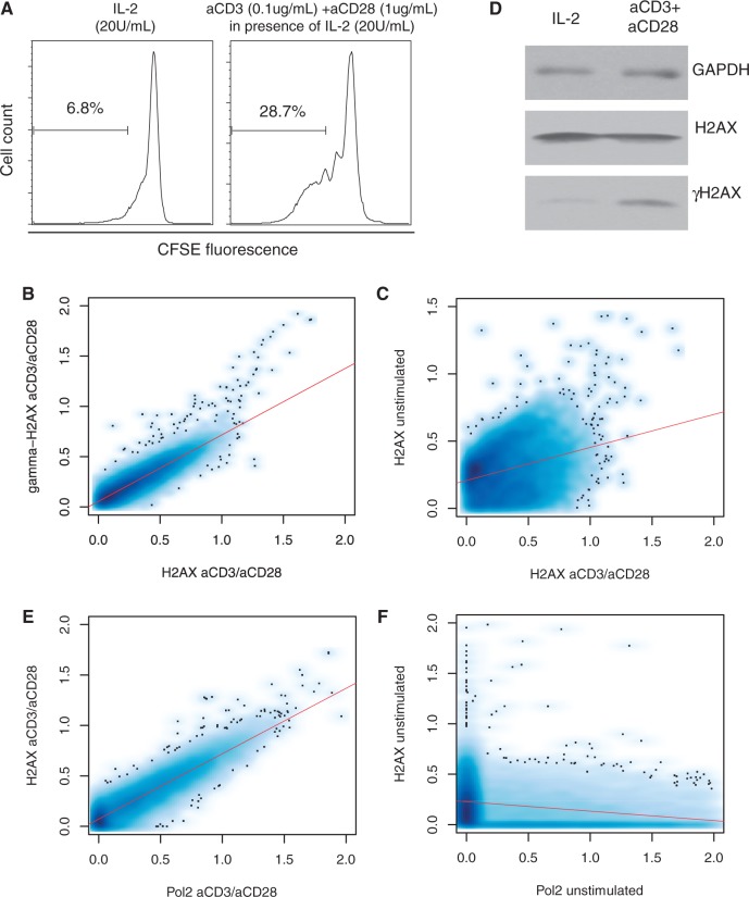
Figure 1.
Genome-wide reorganization of H2AX on cell activation. (A) To assess proliferation of CD4+ T cells, the separated CD4+ T cells were labeled with CFSE. CFSE-labeled CD4+ T cells were re-suspended in 20 U/ml of IL-2. For T cell stimulation, soluble anti-CD3 and anti-CD28 antibodies were added and the culture was maintained for 96 h. (B) Genome-wide correlation between H2AX and γH2AX in the activated T cells. (C) Genome-wide correlation between H2AX in the activated T cells and H2AX in the resting T cells. (D) Western blot of H2AX and γH2AX in IL-2-stimulated cells and cells activated with anti-CD3 and anti-CD28 antibodies. (E) Genome-wide correlation between H2AX and Pol2 in the activated cells. (F) Genome-wide correlation between H2AX and Pol2 in the resting cells.

