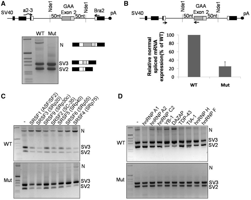
Figure 3.
GAA mRNA splicing variants expressed in cells transfected with the WT and MUT minigenes. (A) Schematic diagram of the minigene system to test the effect of the c.-32-13T>G mutation on exon 2 inclusion. The α-globin, fibronectin EDB and human GAA exon 2 are shown as black, white and gray boxes, respectively. The shaded box represents the segment of exon 2 that becomes included following the activation of the cryptic (c2) acceptor site. Minigenes carrying either WT or MUT GAA sequence were transfected in HeLa cells, and the splicing variants generated were analyzed by RT-PCR using minigene-specific primers (a2-3, Bra2). Cells transfected with the WT construct displayed inclusion of full-length exon 2 (N isoform) as well as total or partial exclusion of exon 2 (SV2 and SV3, respectively). Cells transfected with the MUT minigene expressed SV2 and SV3 variants, while the N variant was undetectable. (B) Relative quantification on the N isoform in cells transfected with WT or MUT minigenes using minigene specific primers designed to amplify only this variant. The abundance of the N isoform generated in cells transfected with the MUT minigene was expressed as percentage of the N isoform detected in cells transfected with the WT minigene. Data represent means ± SD of three independent experiments. (C) The effect of various SR protein overexpressions on exon 2 inclusion was assessed in HeLa cells co-transfected with the WT (upper panel) or MUT (lower panel) minigenes. (D) The effect of various hnRNP protein overexpression on exon 2 inclusion was assessed in HeLa cells co-transfected with the WT (upper panel) or MUT (lower panel) minigenes.

