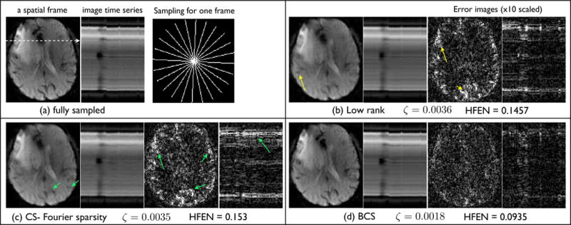
Fig. 8.

Comparisons of the different reconstructions schemes on a brain perfusion MRI dataset. The fully sampled data in (a) is retrospectively undersampled at a high acceleration of 10.66. The radial sampling mask for one frame is shown in (a), subsequent frames had the mask rotated by random angles. We show a spatial frame, the image time series, and the corresponding error images for all the reconstruction schemes. Note from (b,c), the low rank and CS schemes have artifacts in the form of spatiotemporal blur; the various fine features are blurred (see arrows). In contrast, the BCS scheme had crisper features, and superior spatiotemporal fidelity. The reconstruction error and the HFEN error numbers were also considerably less with the BCS scheme.
