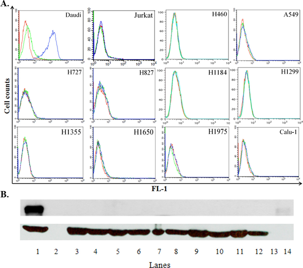
Figure 2. Cell surface CD22 can be detected on CD22+ Daudi cells but not on human lung cancer cell lines.

A. Flow cytometric analysis. One million cells from each cell line were incubated with 25 µg/mL of either a mouse isotype control antibody (MPC-11) or mouse antihuman CD22 (clone HB22.7) MAb. After washing out the unbound primary antibody, a FITC-labeled secondary goat anti-human IgG antibody was used. Cells were analyzed in FL-1 using a BD FACSCalibur. Red line – cells without antibodies; green line – cells plus the isotype control antibody and secondary antibody; blue line – cells plus the anti-CD22 antibody and secondary antibody. This is one representative out of 4–6 independent experiments. B. WB analysis. Equal amounts (25 µg) of protein from cell lysates were subjected to 8% SDS-PAGE followed by a WB with a rabbit anti-human CD22 antibody (H-221 clone). Upper panel - CD22 expression; lower panel - β-actin expression (loading control): 1 – Daudi; 2 - no loading; 3 - Calu-1; 4 - H1975; 5 - H1650; 6- H1355; 7 - H1299; 8 - H1184; 9 - HCC827; 10 - H727; 11 - A549; 12 - H460; 13 - no loading; 14 – molecular weight marker for 140 kDa. This is one representative out of 3–5 independent experiments.
