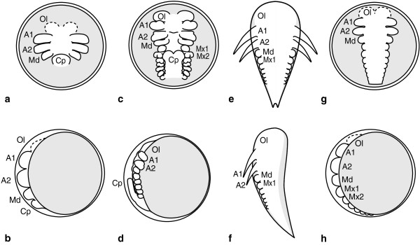
Figure 1.

Schematic overview of malacostracan germ band morphology in embryonic- and pseudodirect development. a, c, e, and g represent ventral views of the germ band, b, d, f and h represent lateral views. a-d Simplified drawings of crayfish embryo modified from [68]. The general pattern applies to all Malacostraca with a yolk-free caudal papilla. a Ventral view of germ band at the egg-nauplius stage. Anlagen of the optic lobes, nauplius appendages and the caudal papilla are present. b Lateral view of same embryo. c Embryo at advanced, but still incomplete germ band stage. Optic lobe- and nauplius appendage rudiments are larger than posterior appendage anlagen. The caudal papilla is flexed anteriorly. d Lateral view of same embryo. e Early mysidacean nauplioid larva. An egg-membrane is missing. First and second antennae show advanced external morphology. Posterior appendage anlagen follow a gradual decrease in differentiation. f Lateral view of nauplioid larva. g Embryo representative of Peracarida except Mysidacea. The gap between naupliar- and postnaupliar appendage development is less distinct. The postnaupliar germ band displays gradual a/p -differentiation. The gradient is exaggerated in this drawing. h Lateral view of same embryo. Areas containing yolk are shaded grey in all drawings. Abbreviations: Ol optic lobes, Cp caudal papilla, A1, A2, Md, Mx1, Mx2, appendage anlagen. Yolk-rich areas are shown in light grey.
