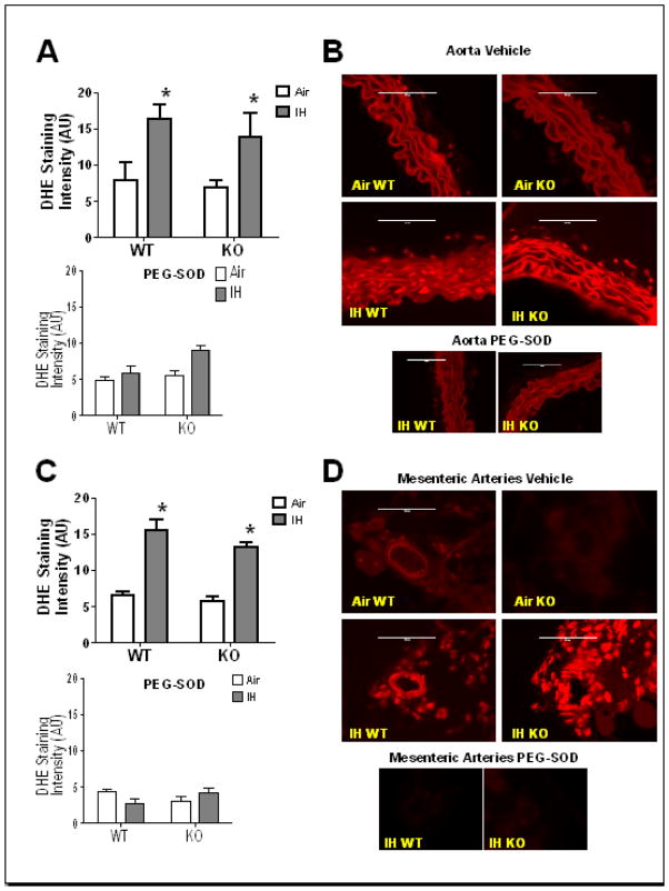
Fig. 1.
IH increases superoxide in aorta (AO) and mesenteric arteries (MA) VSMC. Superoxide levels were measured in discrete regions of interest in the medial layer by DHE staining. Co-incubation with polyethylene glycol conjugated superoxide dismutase (PEG-SOD) reduced fluorescence intensity in IH arteries to or below that of control (Air) arteries. Summary data is shown for A) AO and C) MA of WT and NFATc3 KO mice after 1 week IH or Air exposure, in presence or absence of PEG-SOD. Representative images are shown in B) for AO and D) for MA.*p<0.05 vs. Air for AO; *p<0.01 vs. Air for MA; 2-way ANOVA and Bonferroni post hoc test; n = 4 animals for AO and n = 3 animals for MA. Scale= 100 μm.

