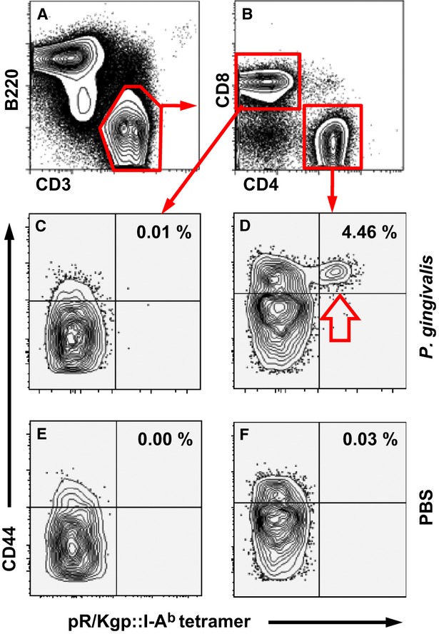
Figure 4.

pR/Kgp::I-Ab tetramer identifies pR/Kgp-specific CD4+ T cells. Mice were orally inoculated and colonized with Porphyromonas gingivalis ATCC 53977 or sham-treated with phosphate-buffered saline (PBS) 21 days before harvesting draining cervical lymph nodes. pR/Kgp-specific CD4+ T cells were enriched using immunomagnetic-positive selection before cell surface staining to discriminate various lymphocyte subsets. (A) Representative FACS contour plots separating B cells (B220+) from T cells (CD3+). (B) CD3+ T cells are sorted into CD8+ (control, C,E) and CD4+ (test D,F) T cells. CD3+ CD4+ and CD3+ CD8+ T cells from P. gingivalis (C,D) and PBS (E,F) orally treated mice. The percentage of pR/Kgp-specific CD4+ T cells found in each sample is given. FACS plots depicted were typical of three independent experiments.
