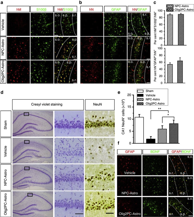
Figure 6. Neuroprotective effects of NPC-Astros and Olig2PC-Astros following GCI.
(a and b) After being transplanted intraventricularly, both Oilg2PC-Astro and NPC-Astro were identified by anti-hN or anti- hM staining in the hippocampal CA1 region. The majority of Olig2PC-Astros and NPC-Astros were also positive for the astroglial markers S100β (a) and GFAP (b). Notably, the GFAP expression by endogenous gliosis in the GCI injury model was suppressed by the transplant astroglia, compared to the vehicle group (right panels). Scale bars represent 20 μm. (c) Quantification of the percentage hM+ S100β+ or hN+GFAP+ in hM+ or hN+ transplanted astroglia, respectively (n=7–8). (d) Representatives of cresyl violet staining and NeuN DAB staining performed on the sections from the hippocampus in the sham group and groups subjected to 14 days of reperfusion after 15 min of GCI with vehicle control and intraventricular administration of NPC-Astros or Olig2-Astros 6 h after ischaemia. The squared areas in cresyl violet staining are shown in high magnification. Correspondent areas were also shown for NeuN staining. Scale bars represent 50 μm. (e) The number of hippocampal CA1 neurons in the different groups measured by stereological analysis (n=6 for each group). (f) Representatives of BDNF expression (green) in the hippocampal CA1 region from vehicle group and groups received astroglial transplants. Astrocytes are labelled by GFAP (red) in the different groups. Scale bars represent 20 μm. One-way ANOVA test; **P<0.01, *P<0.05. Data are presented as mean±s.e.m.

