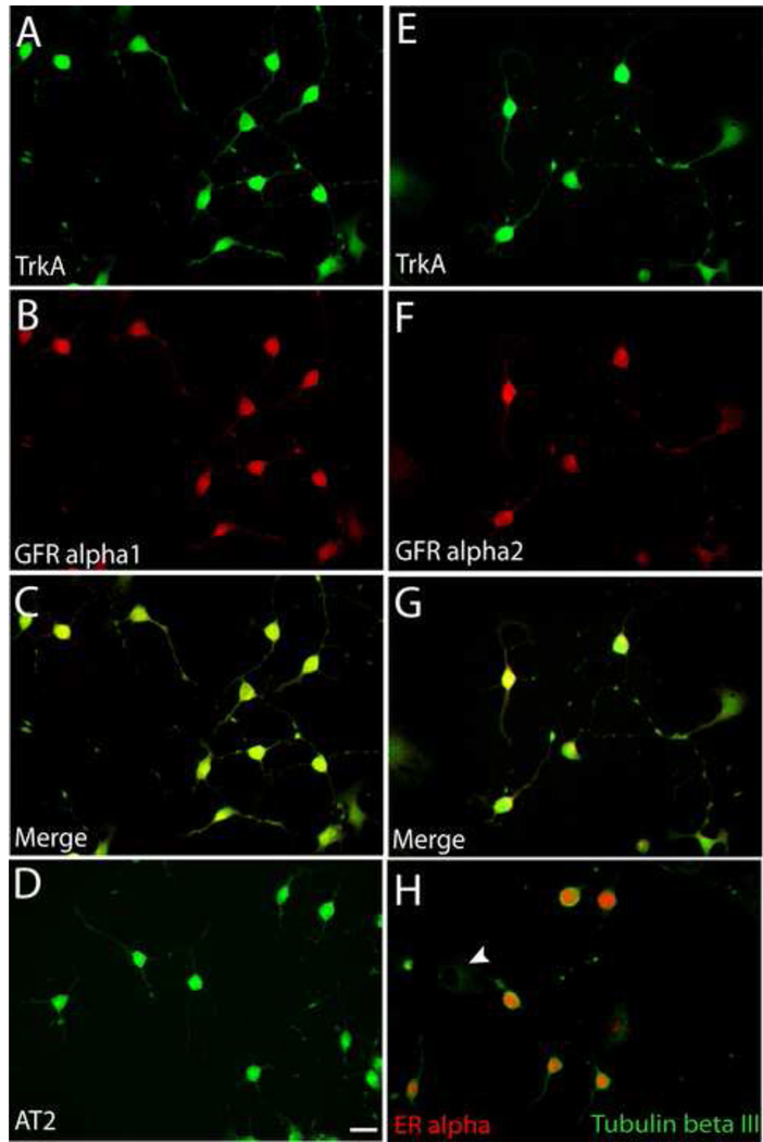
Figure 2.
Expression of growth factor receptors by 50B11 cells. Cells were differentiated for 20h and stained for receptor proteins. A. All cells showing neuron-like morphologies showed immunostaining for the NGF receptor, trkA. B. Cells also showed strong immunoreactivity for the GDNF receptor, GFRα1. C. A merged image shows that all differentiated neuron-like cells express both trkA and GFR alpha1. D. Differentiated cells show immunoreactivity for the ANGII receptor, AT2. E. TrkA staining of differentiated cells. F. Immunostaining of the same field as E shows GFRα2. G. Merged image of E and F shows colocalization of trkA and GFRα2. H. Differentiated tubulin βIII positive cells display predominantly nuclear immunoreactivity for estrogen receptor α whereas undifferentiated cells display little or no ERα. Bar in D =50μm for all panels.

