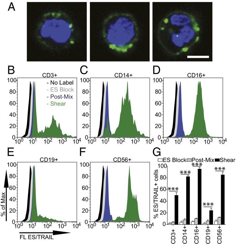
Fig. 2.
ES/TRAIL liposomes adhere to multiple leukocyte subpopulations after exposure to shear flow in whole blood. (A) Confocal images of ES/TRAIL liposomes (green) bound to human leukocytes (blue, cell nuclei) after exposure to shear flow in whole blood in a cone-and-plate viscometer at 188 s−1 for 30 min. Leukocytes have nuclear morphology characteristic of monocytes (Left), lymphocytes (Center), and neutrophils (Right). (Scale bar, 5 μm.) (B–G) To assess the adhesion of ES/TRAIL liposomes to leukocyte subpopulations, fluorescent ES/TRAIL liposomes were added to human blood and exposed to shear flow in a cone-and-plate viscometer at a shear rate of 188 s−1 for 30 min. Leukocytes were isolated from blood using a Polymorphs density gradient and labeled with CD3, CD14, CD16, CD19, and CD56, which is typically expressed on T lymphocytes, monocytes, neutrophils, B-lymphocytes, and natural killer cells, respectively. Expression of fluorescent ES/TRAIL (FL ES/TRAIL) liposomes on the surface of leukocytes that are CD3+ (B), CD14+ (C), CD16+ (D), CD19+ (E), and CD56+ (F), determined using flow cytometry. Expression of CD3, CD14, CD16, CD19, and CD56 on the leukocyte surface was determined using isotype controls. No label, unsheared cells that were not treated with fluorescent ES/TRAIL liposomes; ES Block, cells treated with fluorescent ES/TRAIL liposomes that were pretreated with an ES functional blocking antibody; Post-Mix, cells labeled with fluorescent ES/TRAIL liposomes immediately after mixing liposomes in whole blood. (G) Percent of CD3+, CD14+, CD16+, CD19+, and CD56+ leukocytes adhered to ES/TRAIL liposomes. n = 3 for all samples. Bars represent the mean ± SD in each treatment group. ***P < 0.0001 (one-way ANOVA with Tukey posttest).

