Fig. 1.
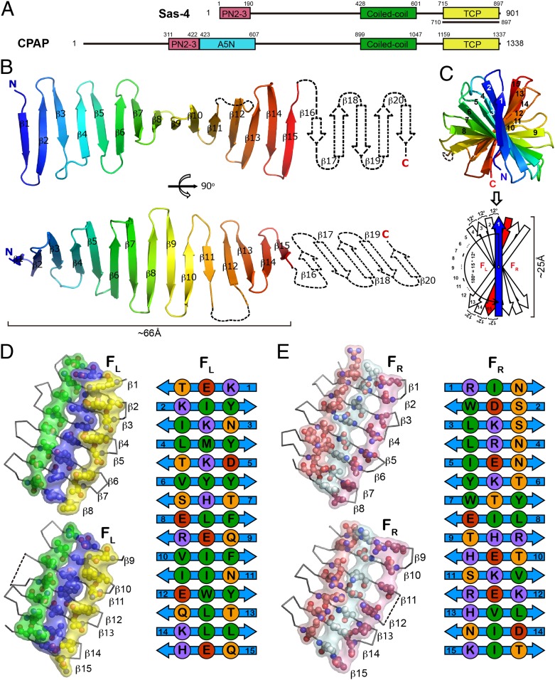
Crystal structure of Drosophila Sas-4–TCP domain. (A) Domain architecture of Drosophila Sas-4 and its human ortholog CPAP. The fragment used for crystallization is indicated by a black underline. (B) Cartoon view of the overall structure of Sas-4–TCP. The invisible part of β16–20 in the crystal structure is shown as dotted lines. (C) Side view of Sas-4–TCP along the longitudinal axis from the N to C termini. Twisting of the TCP β-strands is diagramed below. FL, surface left to β1; FR, surface right to β1. (D and E) Cross-strand ladder residues on FL (D) and FR (E) are shown in spheres and classified into different types by color (purple, positively charged residues; red, negatively charged residues; orange, polar residues; green, hydrophobic residues).

