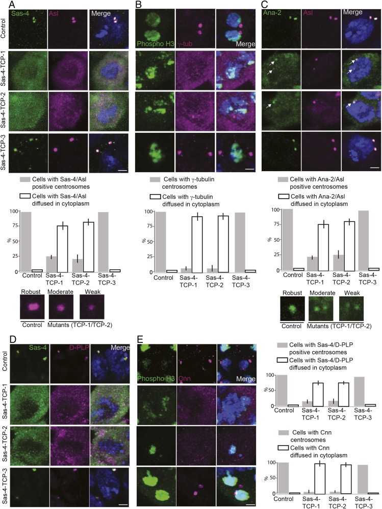
Fig. 4.
TCP domain’s β9–10 surface is essential for tethering the Sas-4–PCM scaffold in a centrosome. (A–E) Spermatogonium centrosomes of control and Sas-4–TCP-3 but not Sas-4–TCP-1 or -2 variants normally recruit Sas-4 (green), Asl (magenta), γ-tubulin (magenta), Ana2 (green), D-PLP (magenta), and Cnn (magenta); ∼75–80% of the cells expressing Sas-4–TCP-1 and -2 variants did not contain detectable centrosomes, and the components of Sas-4 complexes were diffused within the cytoplasm (corresponding graphs for A–E) The remaining cells contained centrosomes but recruited Asl, γ-tubulin, Ana2, D-PLP, or Cnn only at moderate to weak levels compared with control and Sas-4–TCP-3 (Insets shown at the bottom of A and C). The phospho-H3B antibody was used to detect dividing cells for experiments with γ-tubulin and Cnn staining. Arrows in C mark centrosomes. (Scale bar, 1 μm.)

