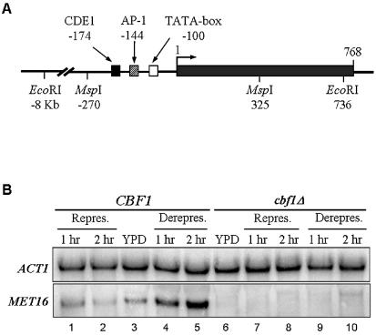
Figure 1.
(A) The MET16 gene structure. The three regulatory elements, CDE1, AP-1 and TATA-box, and relevant restriction enzymes sites are shown. Positions are indicated in relation to the first codon of the protein. (B) Representative results of MET16 transcription in CBF1 (lanes 1–5) and cbf1Δ (lanes 6–10) strains. Hybridization with ACT1 probe (top) and with MET16 (bottom) is shown. Lanes 3 and 6 correspond to samples grown in YPD medium. Lanes 1, 2 and 7, 8 correspond to samples grown for 1 and 2 h in repressing conditions, and lanes 4, 5 and 9, 10 to samples grown in derepressing conditions.

