Fig. 3.
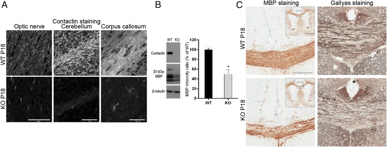
Decreased myelin formation in Cntn1-KO mice. (A) Immunohistochemisty for Contactin on P18 WT or Cntn1-KO (KO) optic nerve, cerebellum, and corpus callosum. Contactin was abolished in KO samples. (Scale bars, 50 μm.) (B) Western blotting of whole-brain samples from P18 WT and KO mice documents the absence of Contactin and ∼50% reduction of MBP in KO samples (levels normalized to β-tubulin, *P < 0.05, n = 3). (C) MBP and myelin (Gallyas) staining of P18 samples shows reduced levels on KO corpus callosum compared with WT. Squares in Insets for MBP staining show the positions of magnified images. (Scale bars, 200 μm; Inset, 1 mm.)

