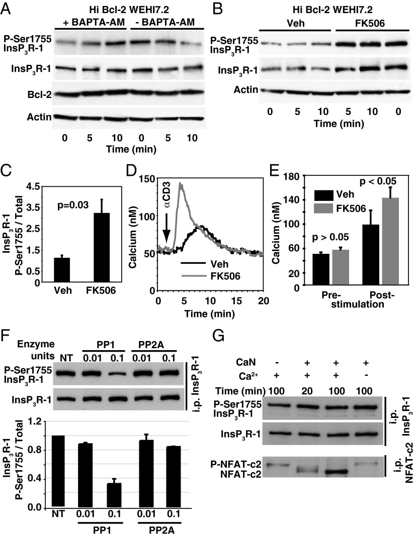
Fig. 2.
CaN and PP1 mediate P-Ser1755 InsP3R-1 regulation by Bcl-2. (A) Immunoblot representative of three experiments indicating that Ca2+ chelation reverses the inhibitory effect of Bcl-2 on anti-CD3 (20 µg/mL)-induced P-Ser1755 InsP3R-1. (B) Pretreatment with FK506 (0.5 µM for 2 h) reverses the inhibitory effect of Bcl-2 on anti-CD3-induced P-Ser1755 InsP3R-1. (C) P-Ser1755 InsP3R-1 levels relative to total InsP3R-1 levels (mean ± SE, n = 3) 5 min after anti-CD3 antibody, indicating that pretreatment with FK506 (0.5 µM for 2 h) reverses the inhibitory effect of Bcl-2 on anti-CD3-induced P-Ser1755 InsP3R-1. (D) Ca2+ traces representing the average cytoplasmic Ca2+ level in 85 cells in individual experiments (n = 3), indicating that pretreatment with FK506 (0.5 µM for 2 h) reverses the inhibitory effect of Bcl-2 on anti-CD3-induced Ca2+ elevation. (E) Summary of cytoplasmic Ca2+ concentrations before and after anti-CD3 addition to Hi Bcl-2 WEHI7.2 cells pretreated for 2 h with DMSO vehicle or FK506 (mean ± SE, n = 3 experiments). (F) In vitro phosphatase assays indicating that PP1, but not PP2A, dephosphorylates P-Ser1755 InsP3R-1 (Upper, representative immunoblots; Lower, P-Ser1755 InsP3R-1 phosphorylation levels relative to total InsP3R-1 levels determined by densitometry of immunoblots; mean ± SD). (G) Immunoblots from a representative in vitro phosphatase assay indicating that CaN does not directly dephosphorylate InsP3R-1 P-Ser1755; Ca2+-dependent dephosphorylation of NFAT-c2 serves as a positive control.

