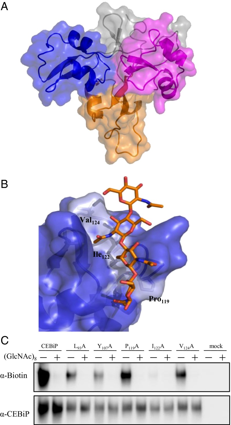
Fig. 4.
Structural basis of chitin oligosaccharides binding. (A) Ribbon and surface representation of the homology model of CEBiP ectodomain. LysM0, LysM1, and LysM2 are drawn in orange, blue, and magenta, respectively. Other regions are colored gray. (B) Modeling of binding of N-acetylchitooligosaccharides to the LysM1domain of CEBiP. (C) Binding of GN8-Bio to mutant CEBiP proteins. CEBiP proteins containing the mutation for the indicated amino acid residues were expressed in N. benthamiana, from which MF for affinity labeling was prepared.

