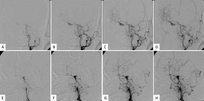
Figure 2.
Series of preoperative digital subtraction angiograms of the left ascending pharyngeal artery (A–D: anteroposterior view; E–H: lateral view) show the feeders were from the branches of the ascending pharyngeal artery and drained via the periclival venous plexus to contralateral sylvian veins and via a posterior fossa vein to the transverse sinus.

