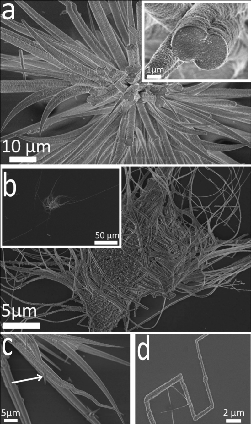
Figure 2.
SEM images of calcite fibers precipitated after 3 days from solutions containing [Ca2+] = 1.5 mM and [PAH] = 0.5 mg mL–1. (a) Fibers growing from a central core, where the inset shows two fibers merging and an internal structure based on nanosized particles. (b) Fibers with aspect ratios of up to 400, (c) a higher magnification image showing straight and convoluted fibers and the formation of branches (arrow) and (d) a fiber showing rapid changes in direction.

