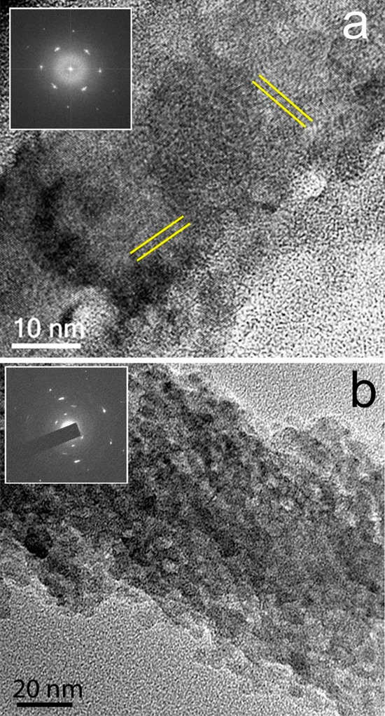
Figure 3.

TEM image and corresponding electron diffraction pattern (insets) of calcite fibers precipitated from reaction solutions containing (a) [Ca2+] = 10 mM and [PAH] = 1 mg mL–1 showing sets of lattice fringes (directions indicated by parallel lines) and (b) [Ca2+] = 10 mM and [PAH] = 1 mg mL–1 showing the nanoparticulate substructure.
