
Figure 5.
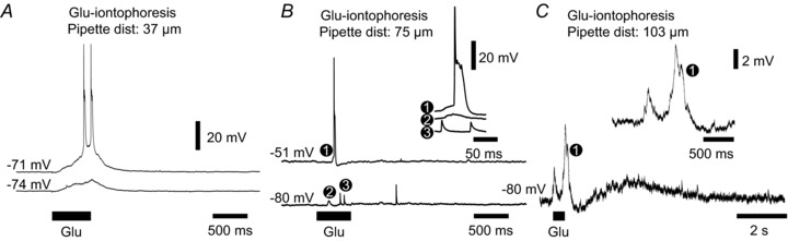
Glutamate iontophoresis induces spikelets and transient depolarizing responses A, whole-cell recording from a DMCC neuron during cluster-inducing glutamate iontophoresis. The neuron was held at rest and at a slightly hyperpolarized level using bias current (lower trace). Note the slowly developing glutamate-induced membrane depolarization during the 500 ms iontophoresis. B, recording from a DMCC neuron during glutamate iontophoresis with a 75 μm inter-pipette distance. Inset: traces at increased resolution corresponding to the full spike at rest (1), the transient depolarization (2) and spikelets (3) recorded at a hyperpolarized level. Note the different spikelet amplitudes during the glutamate iontophoresis. C, recording from a DMCC neuron during glutamate iontophoresis with a 103 μm inter-pipette distance. The neuron was held at subthreshold for spiking during the glutamate iontophoresis. Inset: trace at increased resolution corresponding to the double transient depolarization at (1). Note that the second transient depolarization is followed by a small AP merging into a longer glutamate-induced depolarization.