
Figure 7. Significant overlap between populations of ACh-induced rhythmic inhibitory postsynaptic currents (IPSCs) sensitive to μ-opioid receptor (MOR) and type 1 cannabinoid receptor (CB1R) activation.
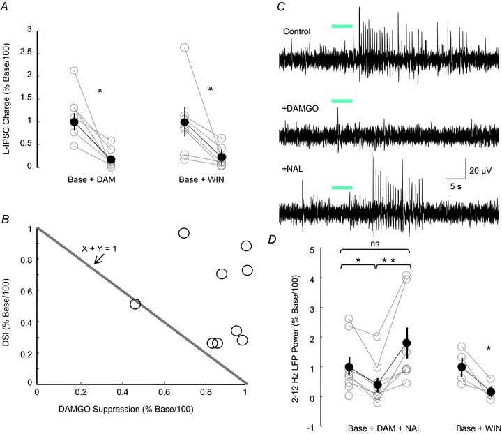
A, group data of the reduction of light-evoked (L-)IPSC charge transfer caused by both DAMGO and WIN55212–2 in ChAT-Cre mice expressing channelrhodopsin2 (ChR2). In two different groups of slices, DAMGO (paired t test, P < 0.05, n= 7) or WIN (paired t test, P < 0.05, n= 7) suppressed the light-induced inhibition by ∼80%, suggesting overlap between MOR-expressing and CB1R-expressing interneuron populations. B, the graph plots the degree of reduction in L-IPSC charge transfer caused by depolarization-induced suppression of inhibition (DSI) versus the degree of reduction caused by DAMGO. The diagonal line is drawn according to x+y= 1, and indicates the locus of points in which the sum of the DAMGO and DSI suppression is linear (on the line), sublinear (to the left of the line) or supralinear (to the right of the line). Data points in the supralinear range are compatible with the conclusion that there is overlap between the populations of MOR-expressing and CB1R-expressing cells generating ACh-induced IPSCs. C, local field potentials (LFPs) induced by release of endogenous ACh are also suppressed by both MOR and CB1R activation. LFP recording from the CA1 pyramidal layer in a slice from a ChAT-Cre mouse expressing ChR2 in cholinergic medial septal axons with ionotropic glutamate receptor (iGluR) antagonists, eserine and 4-aminopyridine (4-AP) present in the artificial cerebrospinal fluid (ACSF). DAMGO reversibly suppresses the light-evoked LFPs. D, as is the case with the ACh-induced IPSCs (B), both DAMGO (left plot, one-way repeated-measures ANOVA, P < 0.001, n= 10) and WIN55212–2 (right plot, paired t test, P < 0.05, n= 4) significantly reduce the LFP power (2–12 Hz).