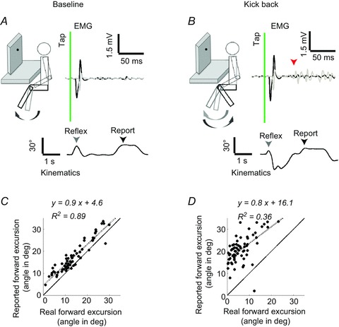
Figure 1. Perception of the knee-jerk reflex in a representative volunteer.

A, schematic side view of the baseline experimental set-up in which the volunteers were seated. They were tapped on the knee and the leg was invisible to the volunteer due to the cardboard baffle. The top traces show single trial EMG recordings from the hamstring (grey) and quadriceps (black). The time of the tap is shown using the vertical line. The angular displacements (lower trace) around the knee joint were documented by using joint angle sensors (in this example) or optical markers. The first arrow on the kinematic trace (grey) depicts the real reflexive movement amplitude and the second marks the reported forward excursion (black). B, when instructed to kick back, a clear activation of the hamstring muscles was seen after the reflexive contraction (arrow – voluntary EMG onset). C and D, scatter plots and linear regressions between the real forward foot movements and the reports, in the baseline (C) or kick back conditions (D). The dashed line shows the linear fit of the displayed data and continuous line depicts the hypothetical ideal observer.
