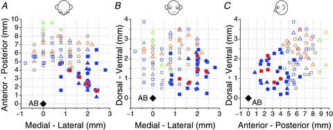
Figure 1. Reconstruction of NU stimulation sites and locations of 31 NU-target VN neurons.

Top (A), frontal (B) and sagittal (C) views showing both NU stimulation sites (open symbols) and NU-target neurons (filled symbols) relative to the abducens nucleus (AB, black diamonds). Note that sites on the left (negative medial–lateral locations beyond 1 mm from midline) were flipped relative to the midline and plotted together with sites on the right (positive medial–lateral locations). Open blue and orange symbols represent NU stimulation sites, which shortened the horizontal VOR time constant (n= 19 and 15, respectively). Open green symbols show four posterior sites whose stimulation elicited nystagmus, without any change in the VOR time constant. Open black symbols represent stimulating sites without effects on the VOR time constant or nystagmus (n= 21). Filled blue symbols show the location of non-EM, NU-target neurons (n= 26), whereas filled red symbols illustrate five NU-target cells that were sensitive to eye movements. All 31 NU-target neurons were identified from stimulation of 19 NU sites with significant behavioural effects (open blue symbols). Different symbols represent neurons/sites from different animals.
