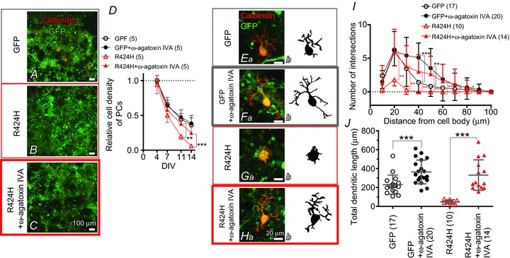
Figure 9. Pharmacological blockade of P/Q-type Ca2+ channels rescues the PC death and dendritic maldevelopment caused by expression of R424H mutant.

A–C, cerebellar cultures expressing GFP alone (A) or R424H mutant with GFP (B and C). The cultures were immunostained for calbindin at DIV 14. In C, ω-agatoxin IVA was added to the culture medium every other day from DIV 2. D, relative cell density of PCs plotted as a function of DIV. The density was normalized to the value of PCs expressing GFP alone at DIV 4. E–H, calbindin-immunolabelled PCs expressing GFP alone (Ea and Fa) or R424H mutant subunits with GFP (Ga and Ha) at DIV 14. Morphologies of PCs are depicted in the right-hand panels for clarity. In F and H, ω-agatoxin IVA was added. I, and J, summary of dendrite complexity measured by Sholl analysis (I) and of total dendritic length (J). *P < 0.05, **P < 0.01, and ***P < 0.001.
