Figure 4. CTAfindings in pulmonary hypertension.
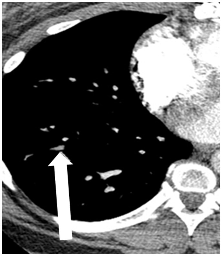
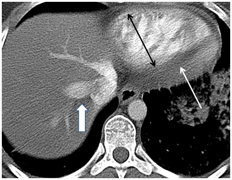
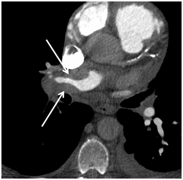
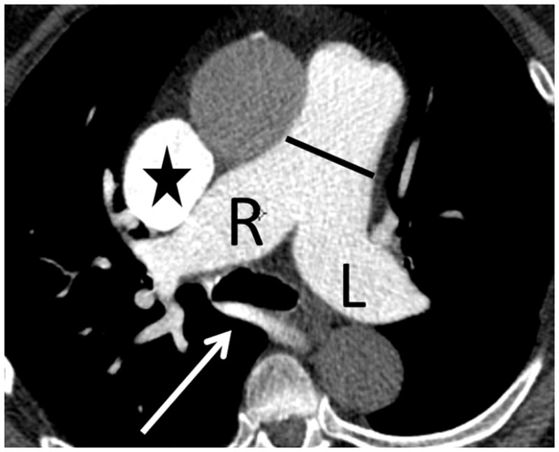
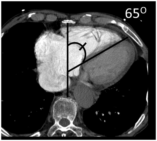
(a) Chronic thromboembolic pulmonary hypertension (CTEPH) with an embolus (arrow) in a subsegmental branch of the lateral segment of the right lower lobe, (b) Elevated pulmonary arterial pressure can be inferred with septal straightening (white arrow) and enlargement of the right ventricle (black arrows). There is reflux into the hepatic veins (white arrow) and inferior vena cava which is an imaging feature of elevated central venous pressure, (c) CTA from a patient with CTEPH showing circumferential chronic clot in the right main pulmonary artery (arrows). This can be removed surgically (pulmonary thromboembolectomy) with a postsurgical lowering of the mean pulmonary arterial pressure. This surgery results in an improved life expectancy for these patients, (d) Dextro- phase CTA showing an enlargement of the superior vena cava (star), pulmonary trunk (black line) and the right (R) and left (L) main pulmonary arteries with reflux into the azygous vein (white arrow), (e) CTA of pulmonary hypertension showing an increased ventricular septal angle at 65 °.
