
Figure 6. 4D flow MRI of Pulmonary arterial hypertension from partial anomalous pulmonary venous return (Dana point 1.4.4).
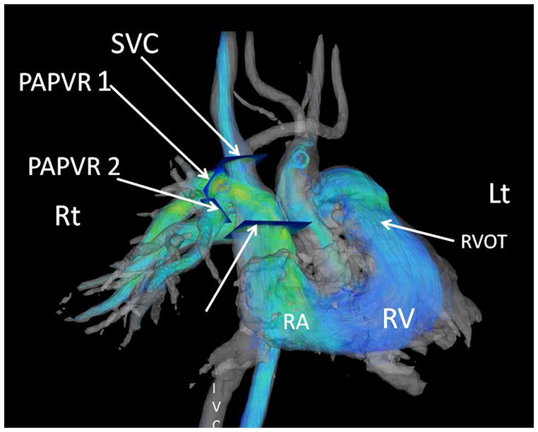
Images were post processed from a respiratory gated Phase Contrast Vastly Undersampled Isotropic Projection Reconstruction (PC-VIPR). These streamlines are color coded for velocity information. The individual contributions of each anomalous vein can be derived from an off line workstation with tools that convert the phase shift information to flow over the cardiac cycle for each manually selected cut-plane. The separate cut planes for the superior vena cava (SVC) and the two anomalous pulmonary veins (PAPVR1 and PAPVR2) are shown by arrows. (Post processing using Encyte™ performed by Phillip Kilgas and Elizabeth Nett, PhD) (Key to abbreviations: IVC- inferior vena cava, RA- right atrium, RVOT- right ventricular outflow tract).