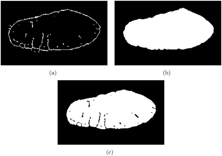
Figure 4.
Segmentation of WGA images stacks. (a) A 3D thinning operator created skeletons of deconvolved images. The skeletons exhibited holes in the outer sarcolemma, which were closed before further processing. (b) Region-growing produced a mask image stack containing intracellular space and t-system. (c) The mask image stack was combined with a thresholded segmentation of the WGA image stack.

